
1. Introduction: The Clinical Utility of EEG
Electroencephalography (EEG) remains one of the most accessible and powerful tools in clinical neurophysiology. In everyday practice, it is often the first test requested when epilepsy is suspected, but its interpretation requires more than recognizing patterns—it requires understanding what those patterns mean in real clinical scenarios.
2. Neurophysiological Basis of the EEG Signal
To interpret an EEG accurately, one must understand the origin of the signal. The scalp EEG does not record individual action potentials. Instead, it captures extracellular dipoles generated by synchronized synaptic activity.
-
The Role of Pyramidal Cells: The pyramidal neurons of the cerebral cortex (specifically in layers III, V, and VI) are aligned perpendicular to the cortical surface. When an excitatory neurotransmitter (e.g., Glutamate) binds to the apical dendrite, an intracellular influx of positive ions occurs, creating an extracellular electronegativity.
-
Spatial Summation: A single dipole is too weak to be detected at the scalp. It requires the synchronous activation of approximately 10 cm² of cortical tissue to generate a voltage measurable by surface electrodes.
-
Volume Conduction: The electrical signal must traverse the pia, CSF, dura, skull, and scalp. This volume conduction attenuates the signal significantly, reducing a 10 millivolt cortical signal to a 10–100 microvolt scalp signal.
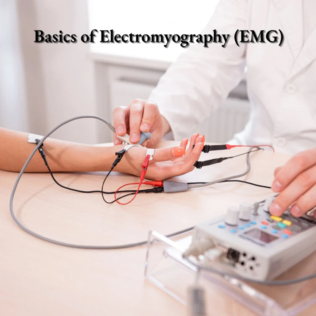
Clinical Pearl: This is a vital distinction in electrodiagnostic medicine. While EMG directly measures highly localized motor unit action potentials (MUAPs) in the peripheral nervous system, scalp EEG relies on massive, synchronized cortical potentials that have been heavily filtered by the skull.
3. Indications for EEG: Evidence-Based Guidelines
The American Academy of Neurology (AAN) and the International League Against Epilepsy (ILAE) provide clear directives on the judicious use of EEG. Over-ordering or misinterpreting EEGs (such as reading normal variants as epileptiform) is a leading cause of epilepsy misdiagnosis.
Primary Indications Include:
-
Diagnosis and Classification of Epilepsy: Differentiating focal onset from generalized onset seizures to guide targeted antiseizure medication (ASM) therapy.
-
Evaluating Altered Mental Status: Identifying non-convulsive status epilepticus (NCSE) or specific encephalopathies (e.g., triphasic waves in hepatic encephalopathy).
-
Prognostication in Anoxic Brain Injury: Following cardiac arrest, specific patterns (e.g., burst suppression, alpha coma) provide critical prognostic data.
-
Surgical Evaluation: In refractory epilepsy, prolonged Video-EEG monitoring is essential for localizing the epileptogenic zone.
4. Normal Background Rhythms and Frequencies
A systematic interpretation always begins with assessing the background rhythm, primarily the Posterior Dominant Rhythm (PDR). Frequencies are categorized into four main bands:
-
Delta (< 4 Hz): Normal in deep sleep (Stage N3) or in infants. In awake adults, focal delta indicates a structural lesion, while generalized delta indicates diffuse encephalopathy.
-
Theta (4 – 7 Hz): Normal during drowsiness or in young children. Can indicate mild dysfunction in awake adults.
-
Alpha (8 – 13 Hz): The hallmark of the awake, relaxed adult with eyes closed. It is maximal in the occipital regions and attenuates (blocks) with eye opening.
-
Beta (> 13 Hz): Fast activity, often prominent frontally. Frequently enhanced by sedative medications, particularly benzodiazepines and barbiturates.
5. Identifying Epileptiform Discharges
According to ILAE definitions, an interictal epileptiform discharge (IED) is a transient waveform that stands out from the background, possessing a sharply contoured morphology.
-
Spikes: Duration of 20 to 70 milliseconds.
-
Sharp Waves: Duration of 70 to 200 milliseconds.
-
Spike-and-Wave Complexes: A spike followed by a slow wave, characteristic of absence epilepsy (classic 3 Hz spike-and-wave) and other generalized syndromes.
Diagnostic Pitfall: A major challenge in clinical practice is distinguishing true IEDs from benign physiological variants (e.g., Wicket spikes, benign epileptiform transients of sleep [BETS], or rhythmic midtemporal theta of drowsiness [RMTD]). Recognizing these mimics is critical to preventing the iatrogenic harm of unnecessary ASM prescriptions.
6. High-Yield Clinical Reasoning: Board-Style Application
Applying neurophysiological concepts to clinical scenarios bridges the gap between theory and practice.
Case Vignette: A 24-year-old medical student experiences a generalized tonic-clonic seizure after two days of severe sleep deprivation. The clinical examination is unremarkable. An awake routine EEG is performed.
Question: What specific activating procedure during the EEG is most likely to provoke the diagnostic abnormality in this patient?
Clinical Logic: Given the patient’s age and the trigger (sleep deprivation), Juvenile Myoclonic Epilepsy (JME) is a primary consideration. In JME, the interictal EEG frequently shows generalized 4–6 Hz polyspike-and-wave discharges. The most potent activating procedure for provoking these discharges in JME is photic stimulation (triggering a photoparoxysmal response), alongside hyperventilation and sleep deprivation itself.
Pingback: 24 Hour EEG at home: What You Need to Know