

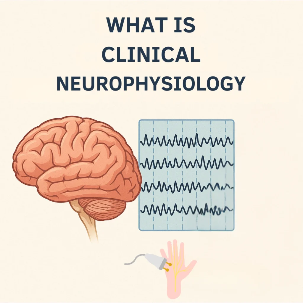
Introduction to EEG
Electroencephalography (EEG) is a non-invasive test that records the brain’s electrical activity. It is one of the most commonly used tools in clinical neurophysiology and plays a vital role in diagnosing and monitoring neurological conditions.
What is EEG?
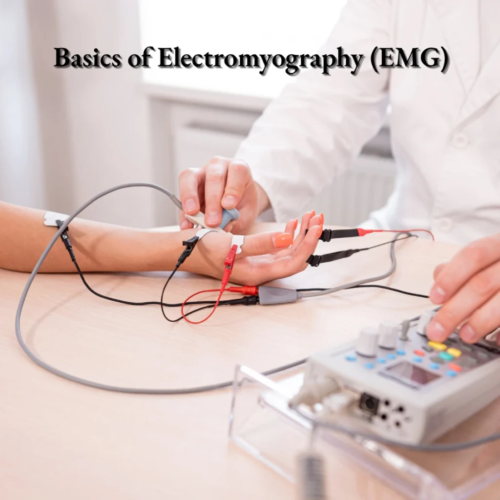
EEG measures the tiny electrical signals produced by neurons in the brain. These signals are captured using small metal discs called electrodes, which are placed on the scalp. The recorded activity is displayed as continuous waveforms on a computer screen.
Why is EEG important?
EEG provides valuable insights into brain function. It is especially useful in:
-
Epilepsy – detecting abnormal discharges and helping classify seizure types.
-
Sleep disorders – studying brain waves during different sleep stages.
-
Brain injury and coma – monitoring brain activity and prognosis.
-
Other neurological conditions – such as encephalitis, metabolic encephalopathies, and unexplained loss of consciousness.
How is EEG performed?
-
Electrodes are placed on the scalp according to the international 10–20 system.
-
The patient is asked to relax, and recordings are taken during wakefulness and sometimes during sleep.
-
Activation procedures (like hyperventilation or photic stimulation) may be used to provoke abnormalities.
Understanding Brain Waves
The EEG records different types of rhythms:
-
Delta (0.5–4 Hz) – deep sleep.
-
Theta (4–7 Hz) – drowsiness or early sleep.
-
Alpha (8–13 Hz) – relaxed wakefulness.
-
Beta (13–30 Hz) – active thinking, alertness.
-
Gamma (>30 Hz) – higher cognitive functions.
Conclusion
EEG remains an essential tool for neurologists and clinical neurophysiologists. Its ability to provide real-time information about brain function makes it invaluable in both research and clinical practice.
Pingback: 24 Hour EEG at home: What You Need to Know